Physical Address
304 North Cardinal St.
Dorchester Center, MA 02124
Physical Address
304 North Cardinal St.
Dorchester Center, MA 02124


The recent discovery of bacterial biofilms within the underlying structure of calcium oxalate kidney stones has led to a major rethinking of the process by which this most common form of nephrolithiasis develops.
Researchers have long known that bacteria such as Proteus Play a role in the development of struvite (MgNH4p4·6H2Q) Kidney stones. However, struvite stones account for less than 30% of all nephrolithiasis cases. Calcium oxalate (CaOx) stones, which constitute the vast majority of cases, have long been thought to be purely inorganic, abiotic, and noninfectious.
New research by biochemist William C. Schmidt and his team at the University of California, Los Angeles, directly challenged this long-held assumption in a recent paper published in the journal Proceedings of the National Academy of Sciences.
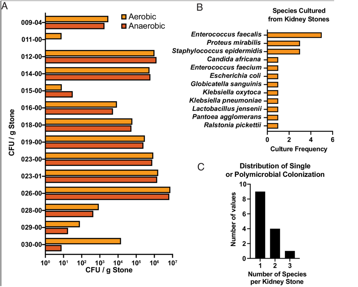
Citing evidence from scanning electron and fluorescence microscopy applied to human CaOx stones, they note that in the internal structure of these stones, bacterial biofilms are “intercalated between polycrystalline mineral layers.” Schmidt and his team observed a similar structure in stone fragments after lithotripsy. Furthermore, DNA analysis showed the presence of bacterial DNA from species including Enterococcus faecalis, Proteus is wonderfuland Escherichia coli – In biofilm materials inside stone fragments.
Furthermore, 17 of the set of 22 CaOx stones they analyzed contained cultivable bacteria. “More than 30% of the stones examined showed some degree of polymicrobial colonization, highlighting the heterogeneity and ecological complexity present in the kidney stone environment.”

The researchers described the results as “unexpected” and wrote that bacteria may play an essential role in the formation of CaOx stones. This could explain, at least in part, why the recurrence rate of CaOx stones is high, and why lithotripsy sometimes leads to kidney infections.
“Our study showed that bacterial presence within kidney stones is widespread across several types of stones, and it is therefore important to consider the possibility of the contribution of bacteria not otherwise detected.”
-William C. Schmidt, UCLA Department of Bioengineering
Schmidt and colleagues point out that the prevalence of kidney stones has risen steadily around the world. A Paper 2023 Estimates are that the lifetime odds are as high as 1 in 11. For some types of stones, recurrence rates are as high as 80%. Taking these trends into account, new insights into etiology may be important for developing better treatments.
The old model of CaOx formation suggests that CaOx crystals begin to form when urine becomes too saturated with calcium oxalate. As these crystals grow, they begin to aggregate, eventually forming structures large enough to obstruct urine flow.
A wide range of substances, including potassium citrate, magnesium, hydroxycitrate, various phosphates, and some urinary proteins such as nephrocalcin, albumin, uromodulin and others, are able to reduce CaOx crystal formation to varying degrees. Some phosphate compounds are actually able to chelate calcium.
The phenomenon of supersaturation is certainly part of the equation, but Schmidt and his colleagues stress that it is not the whole story.
One reason the bacterial agent has been overlooked for so long is the difficulty of culturing bacteria from kidney stones using standard culture techniques. The advanced microscopic and genomic techniques that the UCLA researchers used in this study are not widely available.
Notably, the researchers found evidence of bacterial biofilms in CaOx stones that were culture negative as well as those that were culture positive.
“Our study showed that bacterial presence within kidney stones is widespread across many stone types, and it is therefore important to consider the possibility of the contribution of undetected bacteria that may be present within the stone structure and could contribute to stone-associated infection directly or by stimulating stone growth through mineral deposition and worsening of obstruction.”
How exactly do bacteria affect CaOx kidney stone formation? While researchers are still working to determine the exact mechanisms, bacterial behavior in extreme biochemical conditions holds some clues.
In order to survive, bacteria must maintain a large calcium ion gradient (~100 nM intracellular and ~1 mM extracellular Ca concentration). Pumping calcium out of cells requires a large expenditure of energy, and this metabolic cost is higher when bacteria are in a biofilm mode.
However, organisms in biofilm communities secrete a lot of exopolysaccharides and extracellular DNA, which act as a “sponge” to accumulate extracellular calcium and thus change the gradient and relieve some of the calcium pumping burden.
However, from the point of view of the human kidney, these calcium sponges become a nucleation site for biomineralization of inorganic crystals. In other words, they create seed sites for stone formation.
Schmidt and his colleagues believe their findings have “revolutionary implications” for clinical care. Other than lithotripsy or surgery, there are few treatment options for CaOx kidney stones, and practical prevention strategies are limited to recommendations on dietary changes (reducing sodium, animal protein, and oxalate-rich foods) and increasing hydration. The discovery of bacterial factors in stone formation opens the door to completely new and as yet unrealized possibilities.
“Given the role that biofilm may play in the formation and development of this type of stone, treatments aimed at preventing and eliminating biofilm could have great potential as anti-stone treatment methods in the future.”
This also has implications for preventing recurrence of the disease. Although bacteria may not be detectable by common culture techniques, or appear as nonviable, it is possible that when released from stones during fractionation processing, they become active again and able to grow.
This is clearly a topic worthy of future research.
end